X-linked retinoschisis is a genetic eye disease caused by mutations in the RS1 gene, a gene responsible for making a protein which acts as a kind of "glue" that keeps the layers in the retina together. Because it is an X-linked recessive condition, only males develop the disease, while females are carriers. Without the "glue," the retinal layers begin to separate, causing decreased central vision in both eyes, and up to 20% of patients will experience a retinal detachment at some point during their lives.
Let's look at some images of the retina, first from a normal eye, and then from a patient with X-linked retinoschisis.
Here is a normal configuration of the retina, the inner lining of the back of the eye that gathers light.
Here is an example of the retina in a patient with X-linked retinoschisis. Note the cystic spaces created by the defective retinoschisin protein.
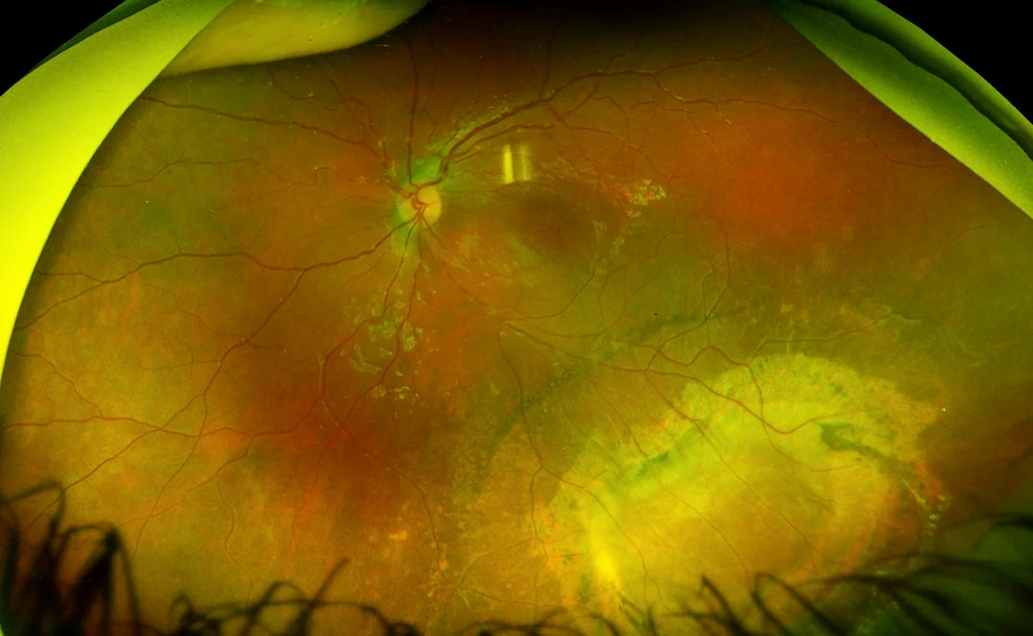
Here is a photo of the retina, showing mild changes in the macula with more extensive scarring and retinal pigment epithelial atrophy in the inferotemporal periphery due to a chronic schisis cavity. The other eye had a very similar lesion.
Carbonic anhydrase inhibitor eyedrops (e.g. dorzolamide) are effective for some patients in reducing the amount of cystic spaces within the retinal layers and in improving visual acuity. Regular eye exams and careful monitoring at home for symptoms of retinal detachment -- such as decreased peripheral vision, floaters, or flashing lights -- are crucial for patients with X-linked retinoschisis.


