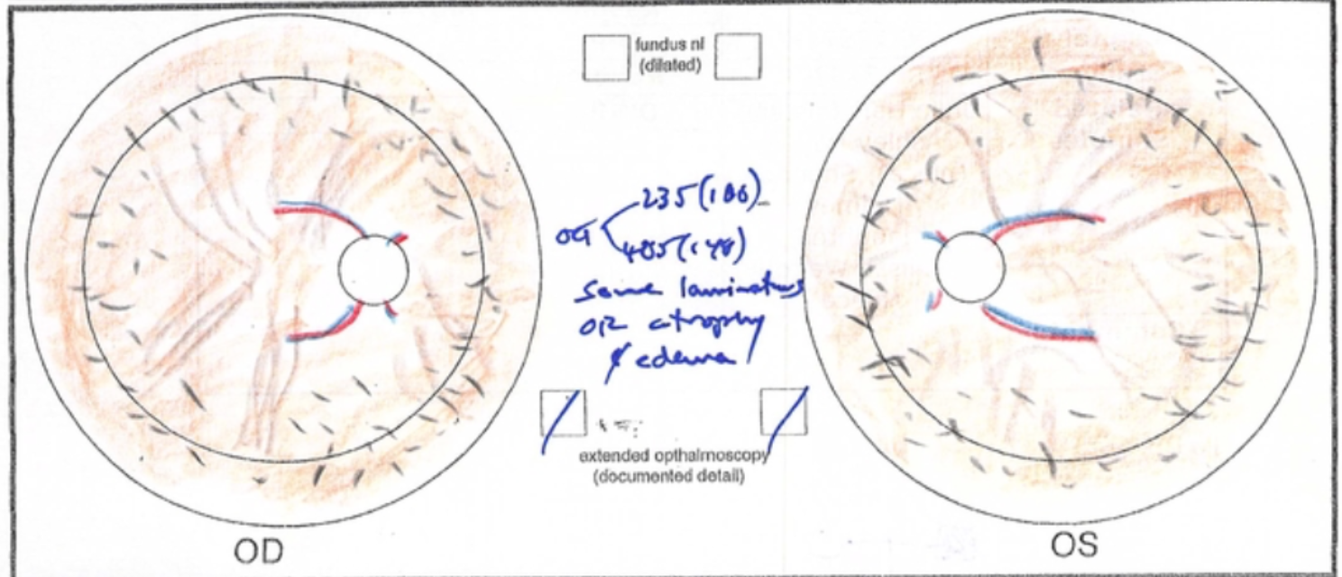
While in training at the University of Iowa's ophthalmology department, I was fortunate enough to have Dr. Stephen R. Russell as one of my mentors. Dr. Russell, a vitreoretinal surgeon and researcher, had himself trained at Iowa, and developed a love for retinal drawings.
In the past, ophthalmologists would often spend 30 minutes or more drawing a picture of what they saw during the patient's eye exam. The retina, the thin layer that lines the inside of the back of your eye, is the most expressive part of the eye, and even the exact same disease can look different from one patient to the next.
Since the advent of improved ophthalmic photographic techniques -- and the increase in the number of patients needing to be seen each day -- ophthalmologists don't take nearly as much time to draw their findings anymore. Lamenting this, Dr. Russell put together a beautiful book containing many exquisite drawings done by dozens of different ophthalmologists at Iowa.
Looking through this book for the first time was an emotional experience. Taking care of people's eyes means so much to me, and I could literally feel the love that these doctors -- all of whom had walked the same halls I was then walking, and many of whom had gone on to become giants in our profession -- had for their patients and their craft as they made these meticulous, beautiful drawings. I decided to put a little more effort into my drawings as time allowed.
Take a look at my own comparatively very unimpressive retinal art in this slideshow, and then scroll down to see a showstopping example from Dr. Russell's book.
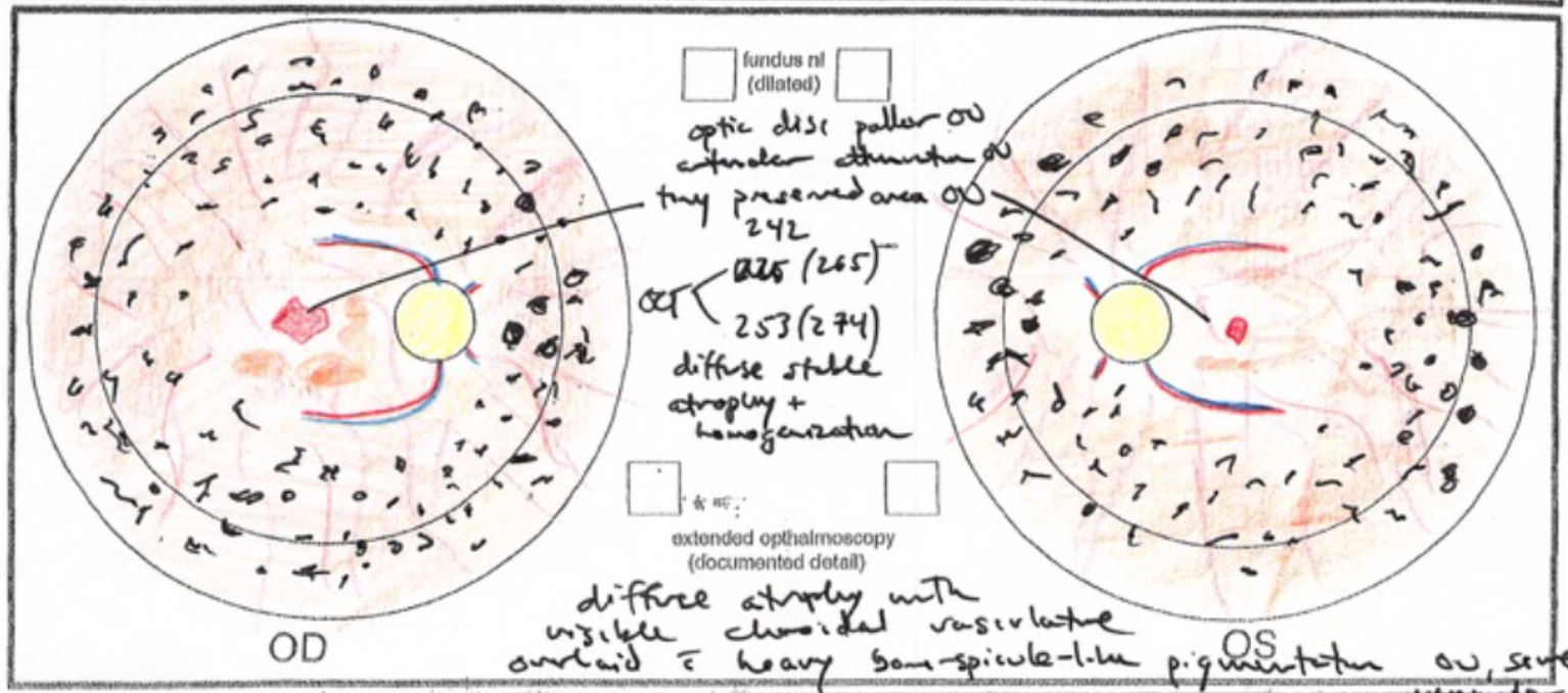
Optic disc pallor, arteriolar attenuation, tiny preserved area of retina within macula, diffuse atrophy with visible choroidal vasculature and heavy bone-spicule-like pigmentation
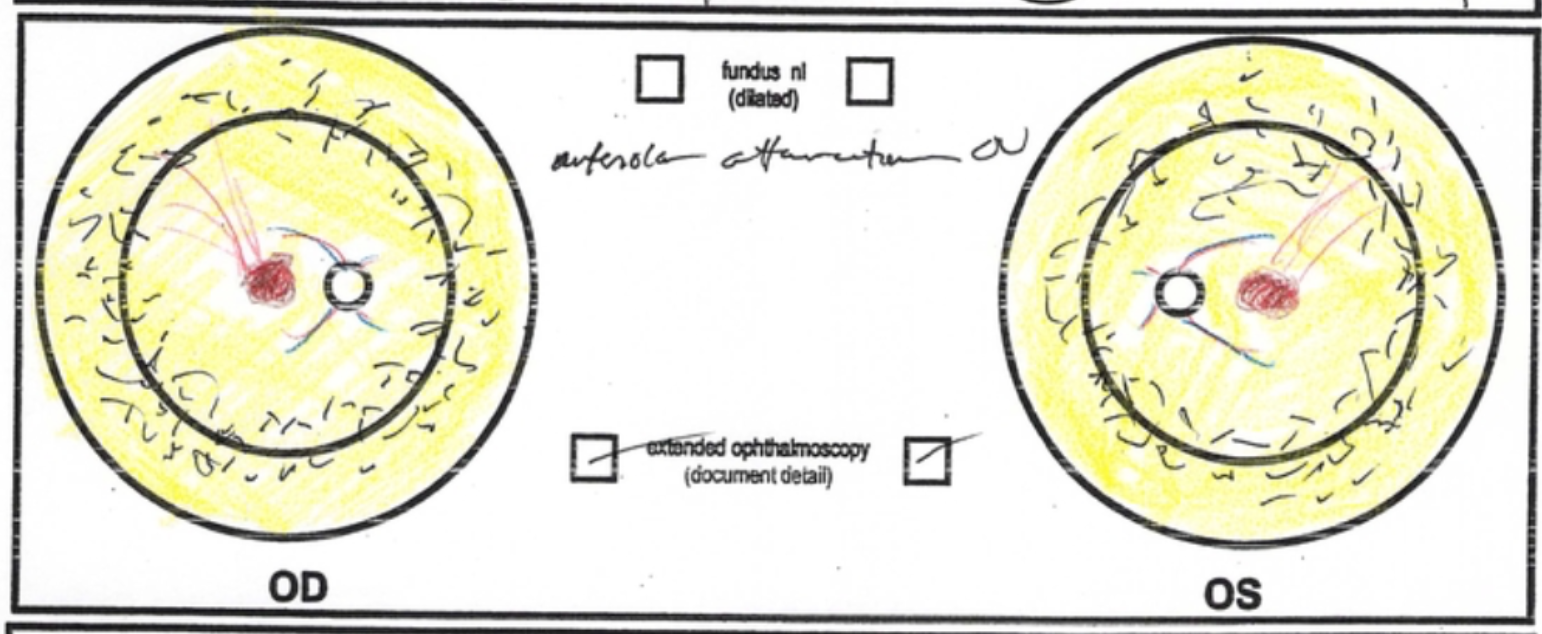
Diffuse arteriolar attenuation, widespread atrophy of retinal pigment epithelium, visible choroidal vasculature
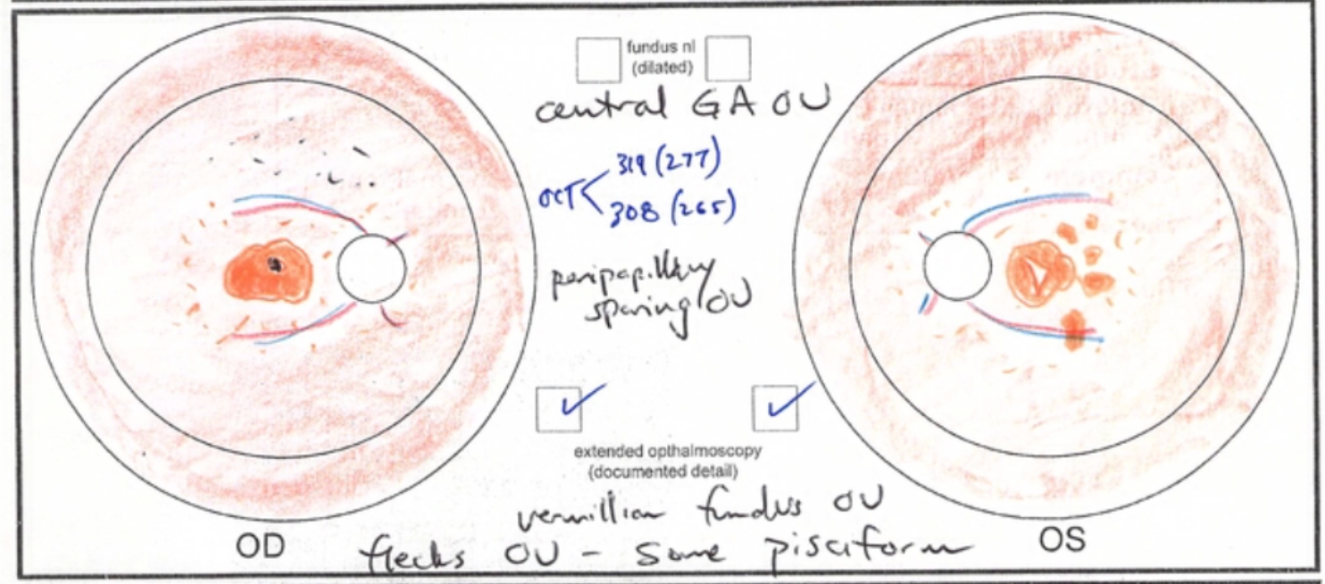
Vermillion fundus, pisciform flecks, central atrophy with overlying pigment, peripapillary sparing
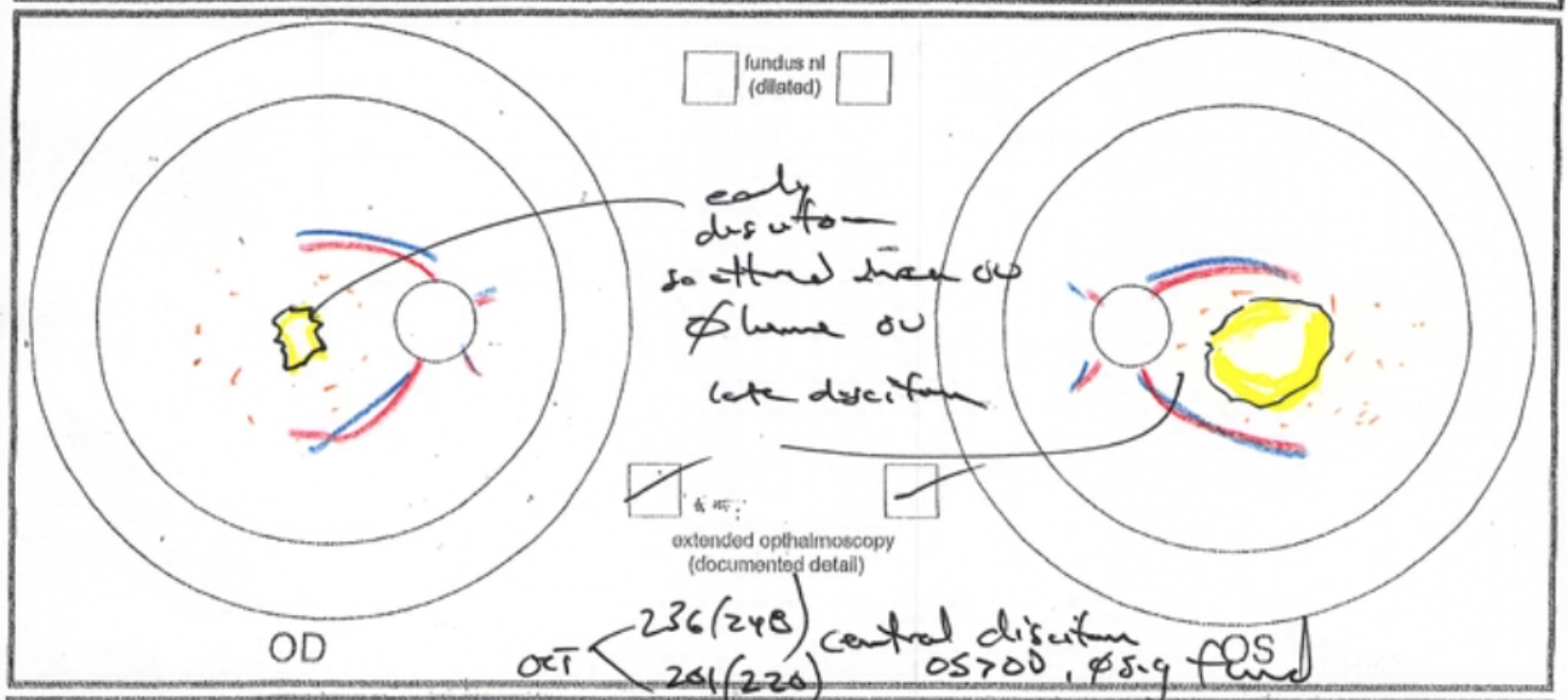
Disciform scars in each eye

Absence of foveal light reflex, with very prominent choroidal vasculature in each eye

Peripheral retinal pigment epithelium atrophy with bone-spicule-like pigmentation, arteriolar attenuation

Central vitelliform (egg-yolk) lesion in both eyes, centered on fovea

Widespread severe atrophy of retinal pigment epithelium with extensive scleral visualization, polygonal black pigment visible on bare sclera

Nummular pigment and atrophy in macula OU with diffuse bone-spicule-like pigmentation; not pictured here are some classic features of CRB1-associated disease: a Coats'-like response, periarteriolar preservation of the retinal pigment epithelium, and disorganized retinal thickening (seen best on OCT)

Hundreds of tiny uniform drusen, clumped in Staphylococci-like clusters


Peripheral retinal atrophy

1) Stable choroidal nevus inferonasally in the right eye; 2) Presumed ocular histoplasmosis syndrome with history of peripapillary choroidal neovascularization in the right eye; 3) Old retinal detachment temporally in the right eye, after laser barricade

Diffuse bone-spicule-like pigmentation in the left eye, combined with just a single patch in the right eye. Initial suspicions were for a somatic RP mutation or AZOOR, but genetic testing revealed a novel RP2 mutation.

Central vitelliform lesions with pseudohypopyon in the left eye. Peripheral lattice degeneration in both eyes.

Hundreds of small tiny drusen in clusters in both maculae

Hundreds of pisciform flecks in each eye, with peripapillary sparing and vermillion fundus

Peripapillary choroidal neovascular membranes (CNVM) in each eye
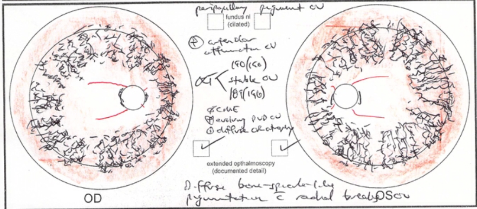
Arteriolar attenuation, diffuse bone-spicule-like pigmentation with radial breaks

Looks like Stargardt disease due to central atrophy and yellow flecks. Very prominent vortex veins.

Diffuse, confluent bone-spicule-like pigmentation, arteriolar attenuation

Preserved central area of retinal pigment epithelium within a sea of atrophy

Central atrophy in each eye with elevated fibrotic area in right eye, macula surrounded by pisciform flecks

Gutters of fluid drain inferiorly in each eye, creating atrophic areas inferior to fovea
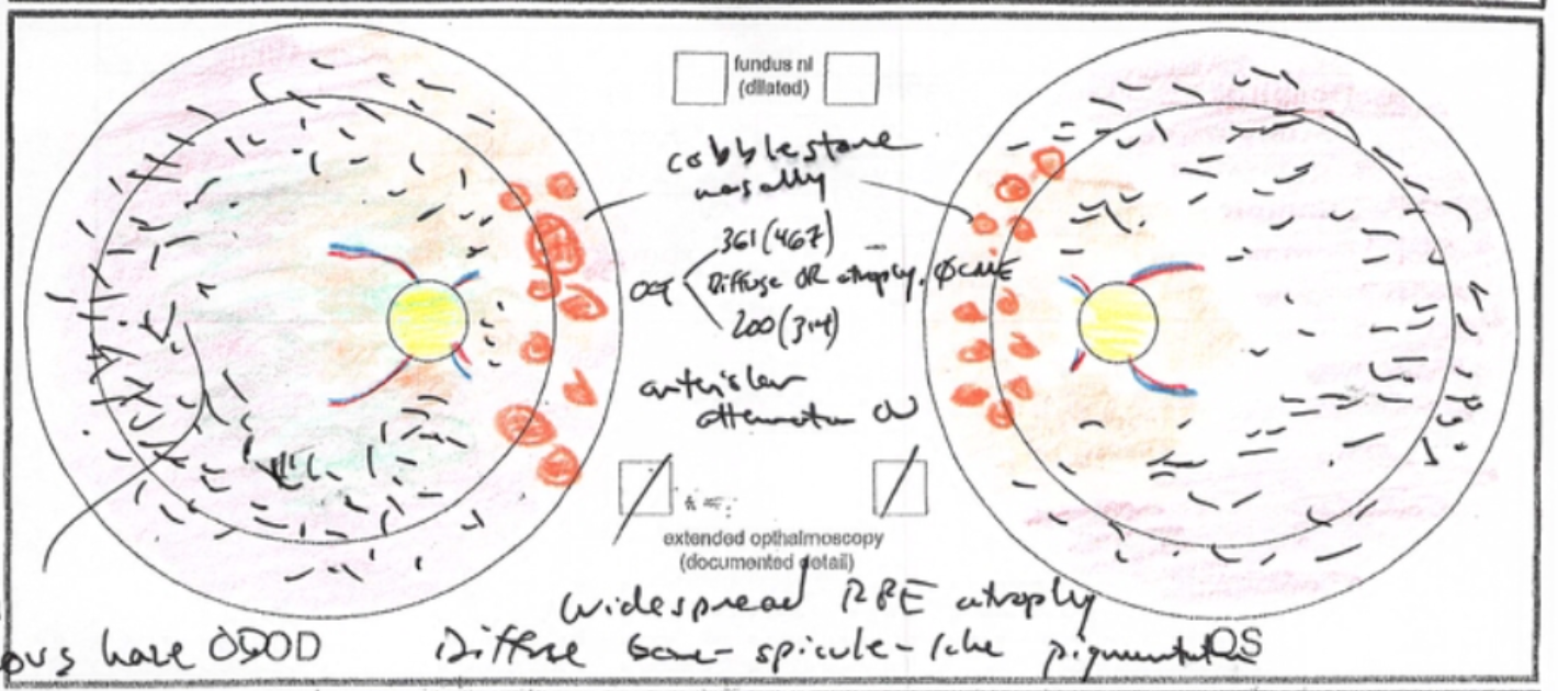
Cobblestone atrophy nasally, diffuse bone-spicule-like pigmentation overlying atrophy of the retinal pigment epithelium
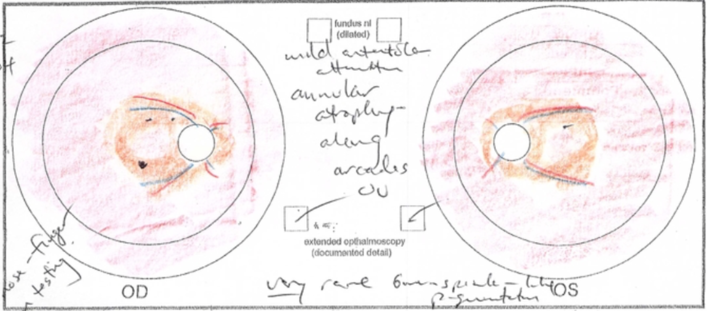
Only rare bone-spicule-like pigmentation, with atrophy along the vascular arcades
