
The optic nerve is a bundle of 1.2 million nerve axons, and is the structure that transmits the visual signal detected by the rods and cones of the retina to the visual cortex in the brain.
This is what a normal optic nerve looks like. You can clearly see where it stops and starts, and it has a healthy pink/yellow hue to it.
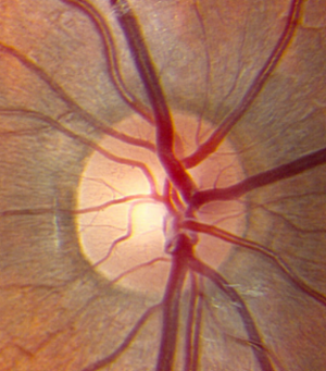
A healthy optic nerve. Image courtesy of eyerounds.org.
The optic nerve can be damaged or abnormal in many ways. In glaucoma, the central, whiter part of the nerve (the "cup") becomes larger as progressive damage occurs due to pressure too high within the eye. The optic nerve can turn pale, meaning it loses its natural pink/yellow hue, because of tumors, nutritional problems, or after lack of blood flow to the nerve, just to name a few.
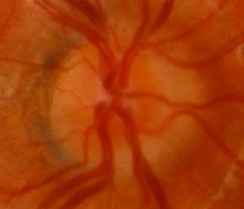
Another way in which the optic nerve can appear abnormal is due to swelling, known as optic disc edema. When the nerve is edematous, the normally crisp edges of the nerve head become blurred and indistinct, almost like someone smudged them with their finger. Take a look at the optic nerve pictured below. There is one large hemorrhage at the 8:30 position, and a number of smaller ones as well. Compare the edges of this nerve with those above; notice how here it's harder to tell where the nerve "starts and stops," so to speak. Finally, examine the blood vessels as they course just outside the optic nerve. Particularly with the two vessels at 4:30 and 5:30, there is an area where it is hard to see the vessels. This is a sign that this nerve is truly edematous, or swollen.

Optic disc edema. Image courtesy of eyerounds.org.
When optic disc edema is due to raised pressure inside the head -- because the nerve is part of the central nervous system, it is bathed with cerebrospinal fluid, and can swell when the pressure inside the skull is too high -- it is known as papilledema. If this is the case, an MRI and MRV (magnetic resonance venogram) should be performed to determine the cause, often followed by a lumbar puncture.
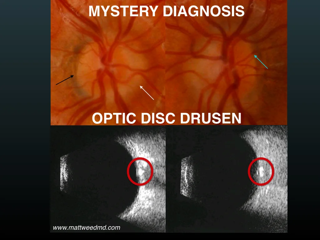
Sometimes, however, the nerve can appear swollen, when in fact it actually isn't. The most common reason? Optic disc drusen. Drusen (from the German word for stone) are small concretions of protein and calcium salts within the optic nerve that may cause the nerve to appear swollen, and are found in 1-2% of the population. While rarely, disc drusen may cause small areas of visual field loss, or even more rarely, sudden vision loss, the vast majority of patients with this condition have zero symptoms.
The diagnosis of optic disc drusen is made with the help of a dilated eye exam and additional imaging, most often an ocular ultrasound. While the pseudo-edema look caused by disc drusen is often present in young children, the drusen themselves often aren't visible to the eye doctor until later in the patient's life. However, many otherwise-invisible drusen can be seen on ultrasound, where calcium within the concretions will show brightly.
In this image below, from a young patient with no symptoms, the black arrow shows a part of the nerve where there is no appearance of swelling. It is easy to see the edge of the nerve. The white arrow shows an indistinct disc margin. The blue arrow shows how it's easy to see these vessels as they enter and exit the nerve (contrast this with the image above), which is a reassuring finding. Encircled in red in the ultrasound images are the disc drusen, which appear as bright white spots.
Thanks to a careful history and examination and the use of a simple ultrasound, which can be done quickly and non-invasively in the eye clinic, the diagnosis of pseudo-edema due to optic disc drusen can be made, and in most cases, further, invasive testing (MRI, spinal tap) can be avoided.
Thank you to The University of Iowa and EyeRounds.org for permission to reproduce this copyrighted material.