FAQs

What is Astigmatism?

This is one of the most common questions parents of my patients ask me in clinic. It’s a word that many of us have heard before, but many people don’t know what it means.

Astigmatism means that the parts of your eyes that focus — the cornea at the very front of the eye and the lens just inside the eye — are not perfectly curved.

In all eyes, images are focused in two dimensions, or axes, onto the retina at the back of our eyes: they are focused vertically and they are focused horizontally. Eyes with astigmatism focus better in one of these directions than in the other. This results in two different “focal points” for the horizontal and vertical directions…which makes your vision blurry.

In the top eye, there is no astigmatism, and the focal points overlap, on the same spot on the retina, so the vision is nice and sharp.In the bottom eye, which has astigmatism, the vertical and horizontal aspects of the image are focused on differen…

In the top eye, there is no astigmatism, and the focal points overlap, on the same spot on the retina, so the vision is nice and sharp.

In the bottom eye, which has astigmatism, the vertical and horizontal aspects of the image are focused on different spots, creating a warped/blurry image.

What are symptoms of astigmatism? Astigmatism may cause blurry vision, either far away or up close or both. It may cause eye strain, headaches, glare, haloes, or visual distortion akin to looking in a “fun house mirror.”

Why does astigmatism not ALWAYS cause decreased vision? Because — dirty little secret alert! — almost all of us have some degree of astigmatism. For the majority, this is minor enough that our vision is still normal, and corrective measures (see below!) are not needed.

What are “normal” levels of astigmatism? That depends on your age. Astigmatism, like its cousins myopia (“nearsightedness”) and hyperopia (“farsightedness”) is measured in units called diopters. Infants and toddlers may have up to two diopters of astigmatism and still be considered normal, and not need glasses. However, as children age, the amount of astigmatism they can tolerate without it affecting their vision declines, and glasses may be required for much less astigmatism.

What causes astigmatism? Astigmatism is almost never the result of anything anyone did or didn’t do, although chronic, frequent eye rubbing can make it worse in some cases. Most often, astigmatism is simply a byproduct of the size and shape of your eyes. Also, just because no one else in your family had astigmatism bad enough to need glasses doesn’t mean you won’t!

What can be done to treat astigmatism? Glasses or contact lenses can be worn to allow the eye to compensate for its astigmatism. Also, contrary to popular belief, laser vision correction surgery, like LASIK, is now excellent at treating astigmatism, even when severe.

What are the consequences of not wearing glasses for astigmatism? In childhood, this may cause permanent developmental delay of the visual system, meaning it may keep a child from ever being able to see normally — even WITH glasses! — in the future. This is called amblyopia. In adulthood, it simply means your vision will be blurry. Astigmatism can get worse over time.

Does wearing an eye patch help fix eye misalignment?

Simply put, no it does not.

This is a common misconception. I frequently see patients who come for a second opinion or because they're new to the area, and their parents tell me something like, "He patched for a few months, but it didn't help, the eye was still crossed in." Patching doesn't help straighten the eyes; it's done to help the non-patched eye develop better vision, but it almost never also results in better eye alignment.

This child has a fun eye patch on his left eye to help his right eye develop better vision. Patching improves eyesight, not eye misalignment.

This child has a fun eye patch on his left eye to help his right eye develop better vision. Patching improves eyesight, not eye misalignment.

Patching treats amblyopia, not strabismus (eye misalignment). Amblyopia occurs when a child's vision doesn't develop normally, and starts to "fall behind." It usually affects just one eye, but rarely may affect both. A child isn't born with the ability to see 20/20; this develops over time. If something is interfering with this process -- for example, eye misalignment -- then the brain often "picks" an eye and develops better vision with that eye, at the expense of the other eye. An analogy I often use is that amblyopia is like having one strong arm and one weak arm: naturally, you want to do everything with your stronger arm, and as you do that, the strong gets stronger and the weak gets weaker.

Patching the better-seeing eye allows the child to rely on their worse-seeing eye, and doing this helps them develop better vision with that eye, sort of like putting the "strong arm" in a sling and doing everything with your weaker arm. This process works best, and quickest, the younger a child is. By age 8-9 years old, a child's visual development is over, and patching is much less likely to be effective.

This can be confusing, because for a child with a misaligned eye that also doesn't see well, while they are wearing their patch on the other eye, the "bad eye" will be straight! This doesn't mean the eye misalignment is better, it just means the child is using that eye. Once the patch comes off, you'll see the eye misalignment again.

As patching continues, a child's vision in the non-patched eye should improve -- they should see better and better on the eye chart (note: preverbal children can certainly also develop amblyopia, and their vision is measured in different ways). However, if they have strabismus, the eye misalignment typically doesn't change much. That doesn't mean patching hasn't worked, as you now know!

For a child with both amblyopia and strabismus, the first step is treating the amblyopia, with some combination of glasses, patching, and/or eyedrops. Once the amblyopia has been treated successfully/maximally, the next step is treating the strabismus, with glasses and/or eye muscle surgery.

To learn more about amblyopia, check out this video from Dr. Weed.

How do I know if my child has pink eye?

Few two-word phrases strike more fear into parents' hearts than "pink eye." This article will help you understand pink eye, what causes it, how it is treated, and when to be concerned.

Pink eye, also known as conjunctivitis, has three common causes:

  1. Viruses

  2. Bacteria

  3. Allergies

Viral conjunctivitis causes red, irritated eyes with mild watery discharge. It often starts in one eye and spreads to the other eye a few days later. Because these same viruses also cause the common cold, patients will usually have either current or recent cold symptoms (cough, runny nose, sore throat, etc.). Viral conjunctivitis, like all viral infections, doesn't respond to antibiotic eyedrops -- you simply have to let it run its course, which can take up to 2 weeks. Artificial tear eyedrops or cool, wet washcloths can be used to soothe the eyes. The best way to prevent this very contagious infection from spreading is frequent handwashing. Also, be careful with commonly-touched objects like towels, doorknobs, toys, phones, etc.

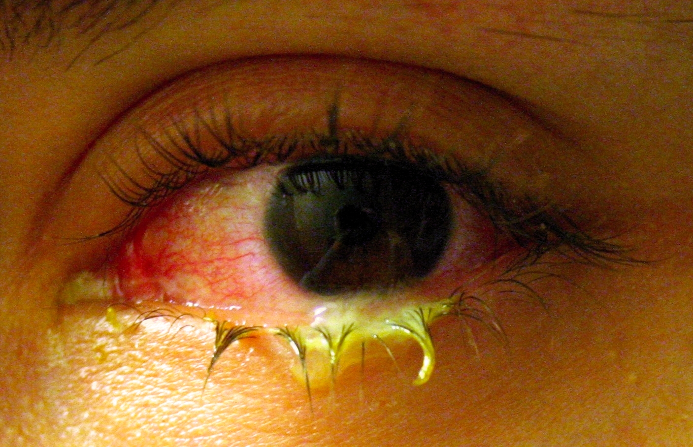
Bacterial conjunctivitis also causes red, irritated eyes, but the discharge is typically thicker, less watery, stickier, and more severe. If your eye doctor suspects bacterial conjunctivitis, you will likely be prescribed an antibiotic eye drop. Most types of bacterial pink eye will resolve after a few days. Like viral infections, bacterial conjunctivitis is also quite contagious, and the same precautions to decreased spread of the infection are important.

The significant, pus-like discharge associated with this conjunctivitis is suggestive of a bacterial infection.

The significant, pus-like discharge associated with this conjunctivitis is suggestive of a bacterial infection.

Allergic conjuncitivitis also causes red, irritated, watery eyes, but the hallmark feature of allergic pink eye is itching. This happens more frequently in people who have asthma, hayfever, or eczema, but it is also common in people who have never had any of these conditions at all. The itching can be intense, and symptom may start very quickly after exposure to the allergen. Young children often have a hard time describing the itching symptom, and they may simply say their eyes hurt or burn. Eye drops, such as the over-the-counter drop ketotifen, can dramatically and quickly reduce symptoms of allergic conjunctivitis.

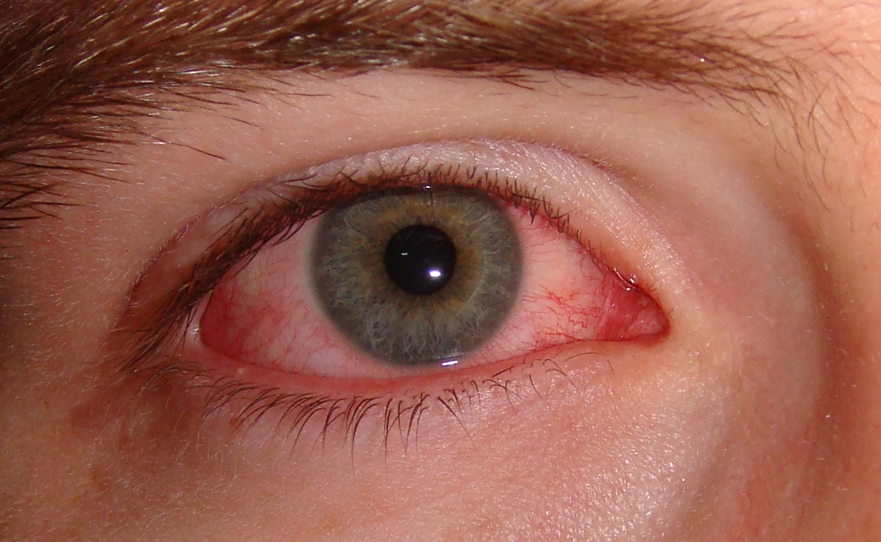
The moderate redness of the eye, combined with the minimal apparent discharge, means this is likely allergic conjunctivitis (if the eyes are significantly itchy) or viral conjunctivitis.

The moderate redness of the eye, combined with the minimal apparent discharge, means this is likely allergic conjunctivitis (if the eyes are significantly itchy) or viral conjunctivitis.

Should I take them to the pediatrician? The urgent care center? The eye doctor? If you're not sure which type of pink eye your child has, take them to see their pediatrician or to an urgent care. If symptoms worsen or persist, take them to see an eye doctor.

Do kids with pink eye need to stay home from school? Not if it's allergic conjunctivitis, since that isn't contagious at all. For bacterial conjunctivitis, children can usually return to school after 1-2 days of antibacterial eye drops. For viral conjunctivitis, most pediatric ophthalmologists recommend keeping kids home until the symptoms resolve, but check your school's policy.

When should I be worried? If you suspect your child may have pink eye, here are some "red flags" that should prompt a call to your eye doctor:

  • Infants, particularly newborns

  • Contact lens wearers

  • Exposure to chemicals

  • Trauma to the eye

  • Prior eye surgery

  • A history of autoimmune disease

  • A visible white "spot" on the eye

  • Symptoms lasting longer than 1-2 weeks

  • Vision loss or severe eye pain

For more information about pink eye, read this article.

Answers to common questions about strabismus (eye muscle) surgery

Here is a short video with the answers to the following eight questions that patients and parents most commonly ask about strabismus surgery.

  1. What are you actually doing in eye muscle surgery?

  2. Is eye muscle surgery done under general anesthesia?

  3. What’s the recovery like after eye muscle surgery?

  4. Will I need to wear a patch after strabismus surgery?

  5. What is the success rate of strabismus surgery?

  6. Is strabismus surgery covered by insurance?

  7. Do you have to take the eyeball out during strabismus surgery?

  8. How old is too old, or too young, to have strabismus surgery?

Can crossed eyes or a wandering "lazy" eye be fixed?

In a word, yes!

People with strabismus -- the medical term for eye misalignment -- whether they have an eye that turns in (esotropia) or wanders out (exotropia), or an eye that goes up (hypertropia), don't have to just "live with it." In almost all cases, they can be treated successfully. The most common treatment options are glasses and eye muscle surgery.

This child has esotropia.

This child has esotropia.

Patients frequently come to see me for strabismus and report having been told by prior physicians, sometimes even eye doctors, that "nothing could be done." I often hear comments after corrective surgery like, "If I had known this could have been fixed, I wouldn't have lived with it all these years!"

Strabismus often causes double vision, especially in adults, and people who haven't lived with double vision rarely grasp how unpleasant, frustrating, and debilitating this condition can be. Even when eye misalignment doesn't cause double vision, in our society, there is an unfortunate but real stigma associated with strabismus, and patients often suffer socially. Children and adults with strabismus are often, incorrectly and unfairly, perceived as less intelligent. Fortunately, as mentioned above, this doesn't need to be the case.

There is no "age limit" to strabismus surgery. This procedure is relatively quick, typically lasting about 40 minutes, and is performed under general anesthesia on an outpatient basis, with patients going home the same day.

Because eye misalignment is often associated with poor vision in childhood, and rarely with an underlying medical problem, if your child's eyes aren't straight, be sure to schedule a visit to see an eye doctor.

To learn more about health insurance coverage for eye muscle surgery, read this.

If you would like to learn about what you might expect in the recovery period after strabismus surgery, click here.

Finally, here is some additional information about reasons why you might consider having strabismus surgery.

Why do I get blind spots with migraine headaches?

I often meet with children in my clinic who report having temporary blind spots in their vision, that seem to grow in size. This can be a frightening experience! Children and their parents are often worried about a severe eye problem in this scenario, particularly if no one in the family has ever experienced similar symptoms.

Fortunately, the eye exam in this setting is almost always normal. How can this be, one might ask? It's because a very common cause of this symptom is migraine, which is a headache syndrome. The root of the problem originates in the brain, not in the eyes -- so the eye exam is normal. The visual symptoms that many people experience with migraine headaches are called "auras."

Migraine headaches are common in children and adolescents, and often follow a course something like this: the person notices a small smudge in their vision, which over the course of a few minutes, expands in size to cover most of their field of vision in both eyes. The edges of this blind spot are often shimmering zigzags, sort of like looking through a kaleidoscope. This aura slowly recedes, and a headache then follows. The headache is often severe, and may be associated with sensitivity to bright lights or loud noises, and the person may be nauseated. Roughly 30% of migraineurs will experience an aura before their headache; sometimes the aura may occur by itself, without a headache.

I recently met a very sharp young patient named Brooke who gave me permission to share her story. She was experiencing frequent blind spots in her vision, and she and her mother were understandably worried by them. I asked if her blind spot started small, like a smudge, and then gradually grew. She nodded. I asked if the edges were shimmering zigzags. She nodded again. I asked if the spot then went away slowly and if she got headaches around the same time. Her eyes got big, she smiled, and said, "You get me!"

To my delight, she then pulled out some drawings that she had made of the vision changes that she gets with her migraine headaches. As a frequent migraineur myself, I found them strikingly accurate. She kindly gave me permission to share them here, so that others could learn about them.

Brooke described how, with her migraine headaches, she would initially get a blind spot with a jagged edge, which a few minutes later would then fill in and be impossible to see through.

Brooke described how, with her migraine headaches, she would initially get a blind spot with a jagged edge, which a few minutes later would then fill in and be impossible to see through.

This is a great example of a jagged, colorful arc that develops at the edge of the expanding blind spot in someone with a migraine aura. It's known as a "scintillating scotoma," or a sparkly blind spot.

This is a great example of a jagged, colorful arc that develops at the edge of the expanding blind spot in someone with a migraine aura. It's known as a "scintillating scotoma," or a sparkly blind spot.

Interestingly, not all auras are visual; some patients experience sensory auras involving a pins-and-needles feeling in the arms or face, which may be followed by numbness.

Patients with visual symptoms followed by headache should consult with their primary doctor. If the primary doctor has concerns about the possibility of a visual problem, a referral to an eye doctor can be pursued.

How often should children have an eye exam?

Parents frequently wonder how often, or when, their children need eye exams. As discussed in this brief video, there are three scenarios in which you should schedule a formal eye exam for your child:

  1. Your child has a symptom or sign of a vision/eye problem, such as blurry vision, an eye that turns in or drifts out, a change in the appearance of the eye, etc.

  2. A physician or teacher is concerned about your child's vision. This is often due to difficulty with a routine vision screening examination.

  3. There is a family history of childhood eye problems.

If you're concerned about a possible eye problem for your son or daughter, schedule an appointment -- I'd be happy to meet you, discuss your concerns, and do a complete eye examination.

What's the recovery like after strabismus surgery?

Strabismus surgery, or eye muscle surgery to realign the eyes, is done on an outpatient basis, so patients are able to go home an hour or two after the surgery is done.

Because strabismus surgery is done under general anesthesia, patients will be fairly groggy afterward, and must have someone with them to drive them home.

There are a few things that are normal symptoms to have after surgery, including:

  • Red eyes, typically lasting a week or two

  • Eye pain, which is usually mild and improves within a few days

  • Blurry vision

  • Eye discharge, which may be yellow, red, or even light green sometimes!

  • Double vision, which is not unusual during the first few weeks of the healing process, especially for adults

Symptoms that are not expected, and should prompt a phone call, include:

  • Severe eye pain not responsive to ice packs or oral pain medication

  • Swelling of the eyelids that is so severe the patient can't open their eyes

  • Worsening redness

  • Vision that continues to get worse

Most patients do very well after strabismus surgery, and need only a few days off from school or work. Plan on seeing me in the office a week after your surgery to check to make sure your eyes are healing normally, and then again six weeks later to assess the outcome of the surgery.