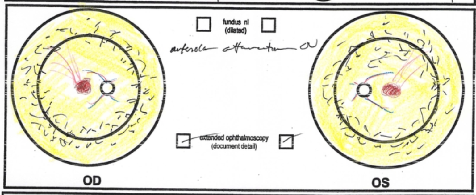
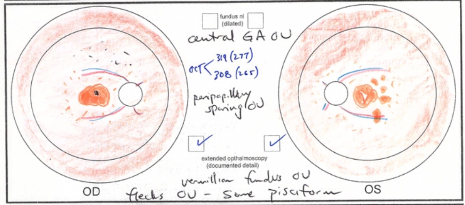
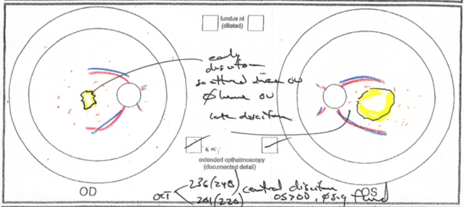
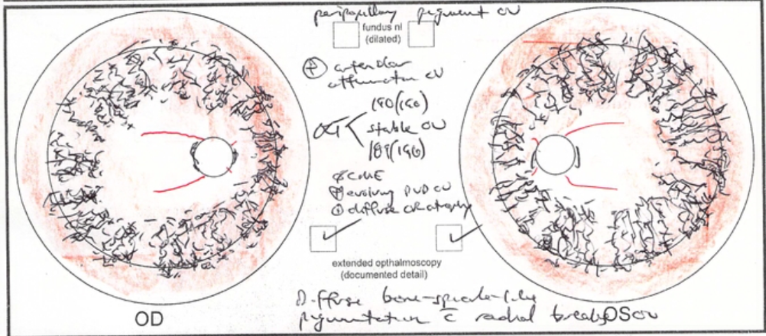
Podcast: Review of Inherited Macular Dystrophies

I was honored to be invited back again as a guest on the Straight From the Cutter's Mouth Podcast this week. This time, host Dr. Jay Sridhar and I discussed one of my favorite subjects: inherited macular dystrophies. It's a succinct review covering the clinical essentials of conditions like Stargardt disease, Best disease, and pattern dystrophy.

Here's a link to subscribe to this top-notch podcast on iTunes.

Mystery Diagnosis: X-Linked Retinoschisis

X-linked retinoschisis is a genetic eye disease caused by mutations in the RS1 gene, a gene responsible for making a protein which acts as a kind of "glue" that keeps the layers in the retina together. Because it is an X-linked recessive condition, only males develop the disease, while females are carriers. Without the "glue," the retinal layers begin to separate, causing decreased central vision in both eyes, and up to 20% of patients will experience a retinal detachment at some point during their lives.

Let's look at some images of the retina, first from a normal eye, and then from a patient with X-linked retinoschisis.

Here is a normal configuration of the retina, the inner lining of the back of the eye that gathers light.

Here is a normal configuration of the retina, the inner lining of the back of the eye that gathers light.

Here is an example of the retina in a patient with X-linked retinoschisis. Note the cystic spaces created by the defective retinoschisin protein.

Here is an example of the retina in a patient with X-linked retinoschisis. Note the cystic spaces created by the defective retinoschisin protein.

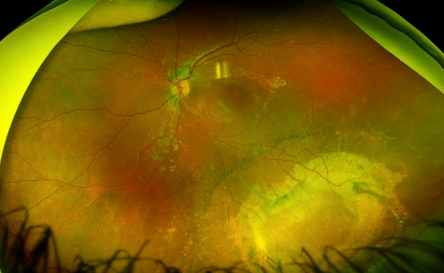
Here is a photo of the retina, showing mild changes in the macula with more extensive scarring and retinal pigment epithelial atrophy in the inferotemporal periphery due to a chronic schisis cavity. The other eye had a very similar lesion.

Here is a photo of the retina, showing mild changes in the macula with more extensive scarring and retinal pigment epithelial atrophy in the inferotemporal periphery due to a chronic schisis cavity. The other eye had a very similar lesion.

Carbonic anhydrase inhibitor eyedrops (e.g. dorzolamide) are effective for some patients in reducing the amount of cystic spaces within the retinal layers and in improving visual acuity. Regular eye exams and careful monitoring at home for symptoms of retinal detachment -- such as decreased peripheral vision, floaters, or flashing lights -- are crucial for patients with X-linked retinoschisis.

Stars' Eyes: Marcelo Lusardi

There are many things in life that look a lot easier than they actually turn out to be once you try them. Surfing. Golf. Writing poetry.

Skateboarding is one of these things. Have you tried it before? Pretty tough, right? Now, imagine doing it blind.

That's exactly what Marcelo Lusardi, a 19-year-old from Santiago de Compostela, Spain, does, ever since he lost vision a few years ago due to Leber hereditary optic neuropathy (LHON).

LHON is a genetic disorder caused by mutations in mitochondrial DNA. Mitochondria, as you may remember from science class, are the "power houses" of our cells, where energy (ATP) is generated, and they contain special DNA which we all inherit exclusively from out mothers. People with LHON typically develop severe painless central vision loss in one eye, often in their teens or 20s.

As if this weren't devastating enough, the other eye usually follows the same course, losing vision a few weeks or months later. This happens in LHON because the energy production in the optic nerves becomes inadequate, causing the ganglion cells within the nerve to die. Although supplementary coenzyme Q-10 (idebenone), combined with avoidance of alcohol and tobacco, may prevent further vision loss, there is at present no way to restore vision for a person who has lost it due to LHON.

Here is a poignant video about Marcelo, entitled "The Blind Rider." My favorite part? 

I used to skateboard. I went blind. I’m still skateboarding. . . . I realized that skateboarding was what I love most in life, and that I was going to keep doing it, come what may.

Ocular Genetics at the Spokane Eye Clinic

At the 2017 meeting of the American Association for Pediatric Ophthalmology and Strabismus, Dr. Alex Levin, pediatric ophthalmologist at Wills Eye Hospital, pointed out that there are only 70-80 ocular geneticists in the world. Dr. Levin himself is one, and so am I. Having completed a year of inherited eye disease fellowship at the University of Iowa with experts like Dr. Edwin Stone, in addition to working as a comprehensive pediatric ophthalmologist and taking care of every type of childhood eye problem, I now dedicate part of my practice to patients with rare, blinding diseases of childhood. I'm happy to be able to offer this service to the region, as area patients formerly had to go to Seattle or Portland to receive this specialty care.

At the Spokane Eye Clinic, my pediatric inherited eye disease clinic takes place once each month, and they are some of my favorite days of work. I schedule just a few patients that day, so that each patient can receive the same high-quality, comprehensive care that I learned to provide in fellowship.

The evaluation begins even before the patients arrive, with a review of the medical record from the patient's referring eye doctor. I read and assess the available information and develop an individualized plan for each patient's visit. Upon arrival, a member of our team initiates the eye exam, and then I visit with each family and take a thorough history, focusing on when the first symptoms began, what they were, how they have changed over time, any prior diagnoses that have been made or genetic testing that has been done, the family history, and several other points.

The Spokane Eye Clinic has built a diagnostic testing suite with specialized equipment that rivals that typically found in an academic medical center.

In fact, the machines we use here are the exact same that I used at the University of Iowa! Some of the advanced technology that I routinely use in the inherited eye disease clinic includes the following:

  • Kinetic visual fields: Whereas standard visual field machines like the Humphrey are designed to check for glaucoma and typically test the patient's central-most vision, the Octopus perimeter allows for the patient's far peripheral vision to be evaluated as well. This is crucial for patients with inherited eye diseases, because these conditions often affect peripheral vision first.
  • Heidelberg Spectralis optical coherence tomography (OCT): The advent of OCT was a revelation within ophthalmology. Equivalent to an ultrasound that uses light instead of sound, OCT allows for detailed imaging of different areas of the eye. This quick, painless test gives a cross-sectional image of the patient's retina, allowing me to see the photoreceptor cells (rods and cones) which are damaged in many inherited eye diseases -- sometimes before the patient has developed any symptoms.
  • Topcon and Optos fundus cameras: These state-of-the-art cameras let our specialized photographers take pictures of the patient's retina, capturing nuances and providing a baseline against which future eye exams may be compared. With the Topcon, our photographers can take several shots and electronically "stitch" them together into a montage image, like you see below. The Optos, an even newer technology, can be a great option to get a quick montage-like image, especially from a wiggly child!
Montage fundus image taken with a Topcon camera

Montage fundus image taken with a Topcon camera

The Diagnosys ERG unit

The Diagnosys ERG unit

  • Electrophysiology equipment, including electroretinography (ERG), multifocal electroretinography (mfERG), and visual evoked potential (VEP). This highly specialized equipment measures the function of the patient's retina and optic nerve. Our ERG and VEP technology is top-of-the-line, and the only setup of its kind in the inland northwest region.

After a detailed history, eye exam, and any diagnostic testing that may be helpful, I review the likely diagnosis with the patient's family and we formulate a plan. This plan could include additional specialized testing, examining family members, or performing genetic testing to identify the change in the patient's DNA that has led to their eye problem. Individualized treatment of patients with inherited eye diseases, with the goal of restoring vision or preventing additional loss, is an exceedingly promising area of research, and dramatic advances have already been made. As additional trials and treatments become available, I will work with the academic medical centers -- like the University of Iowa -- where these are offered, to get my patients "plugged in."

Podcast: Discussing What Went Wrong at the "Stem Cell Clinic"

The folks over at "Straight From the Cutter's Mouth: A Retina Podcast" invited me on today to talk about the article published this week in the New England Journal of Medicine describing three patients who went blind after receiving "stem cell" injections for treatment of macular degeneration. I wrote about this story in greater detail earlier this week here.

I come on at the 23:06 mark, but the entire podcast is well worth your time.

Here's the link to subscribe in iTunes.

Stem Cells and Treatment of Eye Disease

An article published online today in The New England Journal of Medicine has quickly gained recognition within both the scientific community and among the public. The report, published by Ajay E. Kuriyan, MD and colleagues describes three elderly women with macular degeneration who were treated at a so-called "stem cell clinic" in South Florida with the hope that they would regain vision. Tragically, not only did none of the three have any improvement in their vision, but each suffered severe, permanent vision loss as a result. Each went from having vision good enough to drive to being legally blind. 

This clinic, operated by an entity known as U.S. Stem Cell, claims to have treated many patients with all sorts of different ailments (e.g. knee injuries, heart failure, neurological diseases) with "stem cells," which they purport to obtain by removing fat from the patients' own bellies and then purifying this fatty tissue into stem cells (author's note: I have no idea whether they obtained actual stem cells or not, and I'm highly suspicious that whatever they produced wasn't exactly "100% pure," shall we say).

At this center, these cells are then injected into various body parts -- in the macular degeneration patients, directly into the vitreous gel which takes up most of the volume within the eye -- in order to treat the patient's disease.

As an ophthalmologist, I have been asked by patients and friends in the past about similar stem cell clinics, and so I researched them. I quickly realized that what they were selling was scientifically unsound at best, and potentially dangerous at worst, and I advised any who have asked me to stay away and tell their loved ones to do the same. Reading this New England Journal article, I was very sad to see that my concerns were justified.

The three women reported in Dr. Kuriyan's paper not only had both eyes injected under this extremely unvalidated and highly unscientific approach, they had it done to both eyes on the same day, thus exposing both eyes to a risky, unproven therapy -- and they paid $5,000 to do so.

Now, "Who would ever do that?" you might say. And I agree with you, to a point: I certainly never would, and I hope you wouldn't either. But I can understand how it could happen, from a patient's perspective. I dedicate part of my practice to caring for patients with inherited eye diseases, and I see patients with severe vision loss who are desperate for anything to help improve their vision. I regularly speak with family and friends of people who are really struggling with terrible eye diseases, and they so badly want to find something to help. I understand that -- we all feel that way when people we love are hurting.

And this is one of the things that makes me so angry at the people involved in providing this "stem cell treatment." Using pseudoscience (we'll get to this below), they prey upon people's desperation, charge them a not-insignificant sum of money, and offer a solution which any scientist worth his or her salt would immediately recognize as highly suspicious/utter junk.

But that's not the only reason this makes me angry.

As a scientist who has studied and worked and trained with world-leading researchers at the University of Iowa, I believe that stem cell technology -- appropriately developed, studied, and applied -- is an incredibly promising area. But it will not involve sham clinics where patients are treated for all sorts of different diseases with the same "stem cells." It will not involve patients paying money for experimental treatments. It certainly won't involve having both eyes injected on the same day, and the first patients treated will not be patients -- like these three women -- who had useful vision to begin with, and thus, much more to lose.

Let's discuss stem cells for a minute, because stories like these give this technology a bad name and can erode public trust. In 2012, Japanese physician scientist Shinya Yamanaka was awarded the Nobel Prize in Physiology or Medicine for his groundbreaking discovery that adult, mature skin cells could be reprogrammed to turn back into stem cells. These cells are called induced pluripotent stem cells (iPSCs), because they have been induced into becoming cells that can then become a variety of different tissues, like brain, heart, eye, or liver, for example. Think of stem cells as cells that haven't yet decided what they want to be when they grow up.

stemcell.jpeg

Scientists and physicians in all different areas of medicine are excited about iPSCs because of the possibility that a patient's own skin cells could be reprogrammed first into stem cells, and then developed into cells, tissues, and even organs that the same patient could receive as treatment. For example, could we develop new heart muscle cells and transplant them into patients with damage from a prior heart attack?

iPSCs are different from traditional stem cells in two important ways. First, when you think of "stem cells," you probably think of embryonic stem cells, which come from human embryos, and have serious ethical concerns inherent in their use. iPSCs are a totally separate thing -- no embryos are involved at all -- it's just the patient's own skin! Second, because the iPSCs come from the same person who would then receive them as treatment, the cells are immunologically matched, meaning the patient's body won't reject them as foreign, so they won't need to take medicines to suppress their immune system for the rest of their lives.

A number of other cutting-edge technologies could be realistically be coupled with iPSCs to help patients suffering from blinding eye diseases. CRISPR, which is short for clustered regularly interspaced short palindromic repeats, is derived from a bacterial immune system of sorts, whereby bacteria recognize and cut out foreign DNA they have acquired before it can harm them. This same concept can be applied to human cells, allowing for the genome to be edited; using it, a harmful or dysfunctioning gene can be removed, and a corrected version inserted. A patient blind from a genetic eye disease could have stem cells made from their own skin, could have the genetic defect that caused blindness corrected via CRISPR in these cells, could have the cells differentiated into retinal precursor cells, and could then have these cells transplanted into their eye.

And instead of injecting them into the vitreous gel within the eye, and hoping (against all hope) that somehow they make their way where they are supposed to go, ideally, these cells could be placed within a special biopolymer created via a 3D printer that would keep them in perfect alignment and orientation, and then this could be surgically implanted underneath the patient's retina, exactly where the cells belong. 

Researchers at the Wynn Institute for Vision Research at the University of Iowa are pioneering this exciting science. With the help of philanthropic donations, they planned and constructed their own Good Manufacturing Practices laboratory, which is a facility with the highest imaginable standard of cleanliness and sterilization, and the type of laboratory required by the FDA for developing things like iPSCs for transplantation into humans. Here is an article on PubMed describing some of their recent work.

Properly developed and validated, stem cells hold tremendous promise for dramatic advances in medicine. What the charlatans down in Florida have done in this stem cell clinic is akin to someone hearing that "chemotherapy" is a treatment for cancer, and then cooking up some "chemotherapy" in their basement -- without regard for whether the medicine is the right type for the patient's specific type of cancer, whether it is safe, whether it has been tested adequately, etc. etc. etc. -- and telling the patients to bathe in it. Of course it isn't going to work! And not only that, but anyone who hears about it will likely come away thinking "chemotherapy" doesn't work and is really dangerous.

Just like chemotherapy isn't a cure-all, stem cells won't fix everything. Just like chemotherapy, stem cells need to be cautiously studied and judiciously employed. But just like chemotherapy, stems cells hold tremendous promise for treating patients with heretofore untreatable conditions. We should be extremely wary of anyone advertising stem cell treatment at a cost. But we should also realize that stem cells -- namely, iPSCs -- truly represent an area of incredible potential for treating dozens and perhaps hundreds of devastating human diseases safely and effectively.

ReSpectacle: Helping People See

I recently saw a patient in clinic who has been through a tough stretch in life. He has exotropia, which means his eyes drift apart, and this causes him constant double vision, which is why he came to see me. I have also seen his children as patients, and I know his is a family that tries hard, but has many challenges.

During my clinic visit with him, he decided he wanted to have surgery to fix his exotropia, and so we picked a date. I explained that I would need to see him again in clinic once more prior to surgery to remeasure his strabismus to help me with surgical planning, and that for best accuracy, he needed to be wearing glasses; he is significantly nearsighted, and didn't have glasses, so I prescribed him a new pair.

The day of the pre-operative visit arrived, and I entered the room, eager to see him again. Timidly, he let me know that he hadn't been able to get the eyeglasses I had prescribed, because he couldn't afford them. He went on to say that because he couldn't see well without glasses, he wasn't able to drive a car.

Fortunately, most people in the United States are not in the situation where they must choose between being able to see and being able to eat, but many are. This man needed more than just surgery to realign his eyes, and I realized that getting him glasses would likely have an even larger impact than fixing his double vision.

Thinking quickly, I remembered my friend and colleague Dr. Jeffrey Lynch, whom I met while we were both training in ophthalmology at the University of Iowa. Dr. Lynch, recognizing this same issue that faced my patient, started a 501(c)3 nonprofit organization known as ReSpectacle, which, via volunteers, collects high-quality used eyeglasses, cleans them, photographs them, and categorizes them into an online database which patients and providers domestically and internationally can access.

On ReSpectacle's website, the patient or eye doctor enters the patient's glasses prescription, and instantly, the screen displays a number of different possible "matches," complete with a photograph, a description of the glasses, including the prescription, and a color-coded numerical score that describes how well each option matches the patient's own prescription. The user selects the desired glasses, enters a small amount of basic demographic information, and the glasses are shipped for free to the address desired. Amazing!

respectaclesearch

I recently posed a few questions to Dr. Lynch, who kindly agreed to be interviewed for this story.

MW: What prompted you to start ReSpectacle?

JL: A decade ago on an ophthalmic mission trip...I was disappointed with the haphazard organization and poor quality of the used glasses we had available to us using a traditional eyeglass recycling model. There are many people eager to donate their used glasses and there are many people eager to accept them, the challenge lies in effectively transferring the resource.

As I saw it, the problem was two-fold: 1) A glasses prescription is highly specific to an individual (there are over 10 million possible eyeglass prescriptions) 2) An acceptable style frame is often as critical as (or in some cultures more critical than) the accuracy of the prescription.  

A website seemed like an ideal location to store large volumes of donations yet keep them highly searchable and accessible. The inclusion of photographs & other descriptors gives our users the dignity of choosing a preferred style among available options.  

MW: What has been the biggest challenge in starting or running ReSpectacle?

JL: Finding the time to give the organization the attention it deserves, among competing responsibilities and interests as a young physician and parent. 

MW: How do you envision ReSpectacle growing in the future?

JL We expect it to continue growing small chapters organically at academic medical centers, taking advantage of the natural migration patterns of our medical student volunteers as they 'match' to different residency programs across the country. Simultaneously, we will be collaborating and developing our own chapters that can accommodate larger volumes of glasses and offer quicker turnaround times on orders. At some point we expect to grow our network to include international locations as good opportunities arise.

MW: How many pairs of eyeglasses has ReSpectacle recycled?

JL: We recently processed our 10,000th order here in the United States, and have recycled a similar number internationally.

A woman in Nepal with her glasses prescription, which ReSpectacle was able to match and provide. Used with permission.

A woman in Nepal with her glasses prescription, which ReSpectacle was able to match and provide. Used with permission.

MW: Is the process for obtaining glasses internationally any different from doing so within the United States?

JL: Yes, currently we do not support shipping of individual orders internationally, as the cost is prohibitive. Instead, we partner with mission groups or established eye care providers in underserved areas abroad and supply glasses to them in batches. They will typically send us 'mugshots' of patients holding their prescriptions, which are then matched to the best pair of glasses in our database taking into account the prescription power and gender/style. We have worked with over 30 mission groups and offer significant flexibility depending on their needs.

MW: My patient was in disbelief that something like this was even possible, and he was the happiest I have seen him, thanking me with a big smile over and over again. Thanks, Dr. Lynch, for helping him, and thousands of others, enjoy life more fully with better vision.

To learn more about ReSpectacle, including how you can support the cause, click here.

Mystery Diagnosis: Optic Disc Drusen

The optic nerve is a bundle of 1.2 million nerve axons, and is the structure that transmits the visual signal detected by the rods and cones of the retina to the visual cortex in the brain.

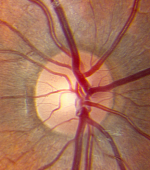
This is what a normal optic nerve looks like. You can clearly see where it stops and starts, and it has a healthy pink/yellow hue to it.

A healthy optic nerve. Image courtesy of eyerounds.org.

A healthy optic nerve. Image courtesy of eyerounds.org.

The optic nerve can be damaged or abnormal in many ways. In glaucoma, the central, whiter part of the nerve (the "cup") becomes larger as progressive damage occurs due to pressure too high within the eye. The optic nerve can turn pale, meaning it loses its natural pink/yellow hue, because of tumors, nutritional problems, or after lack of blood flow to the nerve, just to name a few.

Another way in which the optic nerve can appear abnormal is due to swelling, known as optic disc edema. When the nerve is edematous, the normally crisp edges of the nerve head become blurred and indistinct, almost like someone smudged them with their finger. Take a look at the optic nerve pictured below. There is one large hemorrhage at the 8:30 position, and a number of smaller ones as well. Compare the edges of this nerve with those above; notice how here it's harder to tell where the nerve "starts and stops," so to speak. Finally, examine the blood vessels as they course just outside the optic nerve. Particularly with the two vessels at 4:30 and 5:30, there is an area where it is hard to see the vessels. This is a sign that this nerve is truly edematous, or swollen.

Optic disc edema. Image courtesy of eyerounds.org.

Optic disc edema. Image courtesy of eyerounds.org.

When optic disc edema is due to raised pressure inside the head -- because the nerve is part of the central nervous system, it is bathed with cerebrospinal fluid, and can swell when the pressure inside the skull is too high -- it is known as papilledema. If this is the case, an MRI and MRV (magnetic resonance venogram) should be performed to determine the cause, often followed by a lumbar puncture.

Sometimes, however, the nerve can appear swollen, when in fact it actually isn't. The most common reason? Optic disc drusen. Drusen (from the German word for stone) are small concretions of protein and calcium salts within the optic nerve that may cause the nerve to appear swollen, and are found in 1-2% of the population. While rarely, disc drusen may cause small areas of visual field loss, or even more rarely, sudden vision loss, the vast majority of patients with this condition have zero symptoms.

The diagnosis of optic disc drusen is made with the help of a dilated eye exam and additional imaging, most often an ocular ultrasound. While the pseudo-edema look caused by disc drusen is often present in young children, the drusen themselves often aren't visible to the eye doctor until later in the patient's life. However, many otherwise-invisible drusen can be seen on ultrasound, where calcium within the concretions will show brightly.

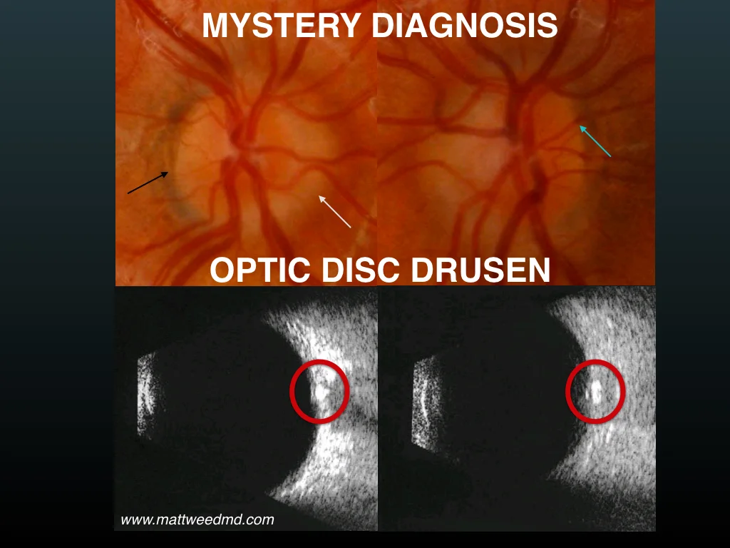
In this image below, from a young patient with no symptoms, the black arrow shows a part of the nerve where there is no appearance of swelling. It is easy to see the edge of the nerve. The white arrow shows an indistinct disc margin. The blue arrow shows how it's easy to see these vessels as they enter and exit the nerve (contrast this with the image above), which is a reassuring finding. Encircled in red in the ultrasound images are the disc drusen, which appear as bright white spots.

Thanks to a careful history and examination and the use of a simple ultrasound, which can be done quickly and non-invasively in the eye clinic, the diagnosis of pseudo-edema due to optic disc drusen can be made, and in most cases, further, invasive testing (MRI, spinal tap) can be avoided.

opticdiscdrusen

Thank you to The University of Iowa and EyeRounds.org for permission to reproduce this copyrighted material.

Stars' Eyes: Abraham Lincoln

Widely regarded as perhaps the greatest of the Presidents of the United States of America, Abraham Lincoln held office during the Civil War, from 1861 until his assassination in 1865. Many aspects of Lincoln's life -- from his humble beginnings in Kentucky and Indiana, to his practicing law before becoming a leader in the nascent Republican party -- are well known. But did you know that he also had strabismus?

lincoln strabismus

Look carefully at this image. Notice how his left eye appears to be looking higher than his right. This is suggestive of a vertical strabismus (eye misalignment), as are the reports that Lincoln suffered from double vision and that his left eye would "roll upward when he was excited or tired."

The intermittent nature of this symptom is consistent with either an intermittent left hypertropia or a dissociated vertical deviation. Both of these conditions are easily diagnosed by a pediatric ophthalmologist, and treatment options may include prisms glasses or corrective surgery.

Because of his very tall, thin stature, many historians have suggested that Lincoln had a condition called Marfan syndrome, a genetic disease which affects the connective tissues in the body. People with Marfan syndrome are typically very tall, thin, and "loose jointed." They are more likely to have strabismus, and may also develop early cataracts, glaucoma, corneal problems, or retinal detachments.

Note: Special credit to my partner, Dr. Jeffrey Colburn MD, of the Spokane Eye Clinic, for making me aware of President Lincoln's strabismus.

PEDIG Meeting -- Tampa, Florida

This week, I've had the opportunity to attend the annual meeting of the Pediatric Eye Disease Investigator Group (PEDIG) in Tampa, Florida. PEDIG, founded in 1997 and funded by the National Eye Institute/National Institutes of Health, is a collaborative network dedicated to supporting research in amblyopia, strabismus, and other childhood eye disorders.

PEDIG

As part of my practice at the Spokane Eye Clinic, my partner Dr. Colburn and I are honored to participate in several PEDIG research studies. This research allows us to help define and develop new and cutting-edge treatments for children with eye problems.

Prior PEDIG research has helped us learn these important findings, among many others:

  • Infantile esotropia: Constant, large esotropia (eye crossing) in infants is exceedingly unlikely to go away on its own, and very likely to require corrective surgery.
  • Treatment of amblyopia: For moderate amblyopia, with vision in the 20/40 to 20/100 range, part time patching and atropine eye drops are equally effective treatments.
  • Nasolacrimal duct obstruction: For infants between 6 and 10 months old who have tear duct obstruction, there is a 66% chance the symptoms resolve without surgery over the next 6 months.

To see the complete list of over 100 PEDIG research articles published in peer-reviewed scientific journals, click here.

Although I can't discuss the actual research data I've seen at this meeting -- stay tuned, as it will be published in scientific journals in the near future! -- I can say that there are a number of exciting potential advances in treatment of retinopathy of prematurity, amblyopia ("lazy eye"), and strabismus (eye misalignment).

One wonderful thing about PEDIG is that it allows eye doctors in private practice, like myself, to participate in nationwide research studies along with physicians in university settings. I am grateful for the chance to help both current patients and future generations by learning more about childhood eye disease and how best to treat it. I've enjoyed the chance to reconnect with old colleagues and make friends with a lot of highly intelligent, motivated people, all of whom want the same thing I do: to figure out how best to take care of our little patients and their eyes.

Guest on "Straight From the Cutter's Mouth" Podcast

I had the great honor of being invited on the "Straight From the Cutter's Mouth" podcast this past week. The host, Dr. Jayanth Sridhar MD, is a friend and colleague whom I met while we were both interviewing for ophthalmology residency positions.

We talked, among other things, about taking care of patients with inherited eye diseases like retinitis pigmentosa and Stargardt disease, as well as strabismus in patients with history of scleral buckle surgery to correct a retinal detachment. We had a great discussion, and I hope you like it!

The episode can be found here, and you can subscribe to the podcast on iTunes here.

Through Their Eyes

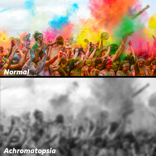
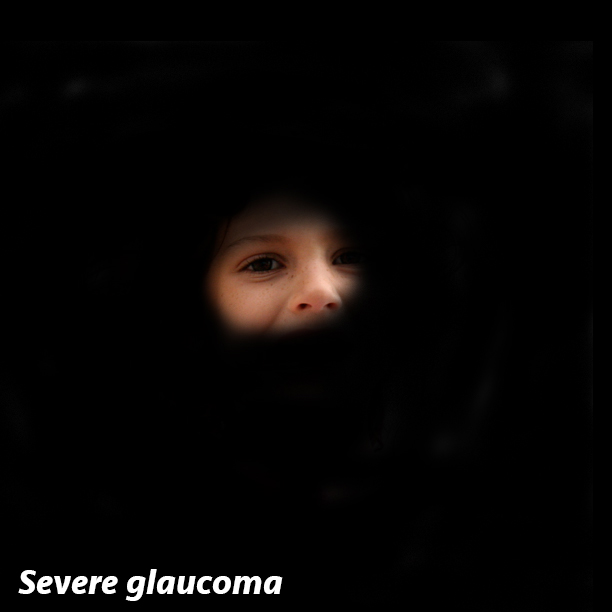
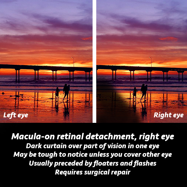
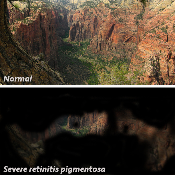
Different vision problems can affect our eyesight in different ways; not all eye problems cause blurriness or can be fixed with glasses. Here are some images I created that help illustrate how the world looks with different types of eye conditions. These images are based on the pathology that is causing the issue and on my experiences talking with patients.

How to Be Successful When on Call

I was on call over Christmas here in Spokane, and it was fairly busy. Not as busy as it would be for some other specialties, but still busy. For 96 straight hours, I was the ophthalmologist covering all consults for three hospitals in the city, and I was also on call for the patients in our call group, which consists of about 25 ophthalmologists. 

For the past eight years, I've had the opportunity to be on call hundreds of times. It's not my favorite part of my job -- and I'm not sure it is for anyone! -- but I've found a few tips that have helped me be successful on call:

1. Manage your expectations. If you assume you won't be busy, at best you will be satisfied, and more than likely you will be disappointed. By contrast, if you prepare for nonstop action, you will be pleasantly surprised and grateful if you get some sleep or time to sit down.

2. Be efficient. Focus on the "W-I-N" strategy: what's important now? What do you need to do to save this person's life/limb/vision until the morning? Don't get bogged down.

3. Most importantly: Remember, you signed up for this. Your patient did not. He or she is seeing you at one of the most vulnerable times in their life. They need your help.

When I posted this on my Facebook page, Dr. Tom Oetting, a great mentor and friend, had the following to say:
People are scared and often just need reassurance. Took me a long time (and having kids) to figure out just how anxious we as parents can get. . . Even though some of the things we see on call seem trivial to us they can really jazz up a family unit! So we shouldn't be surprised or resentful if some of our on call activity seems trivial.

Another friend, the retina surgeon Dr. Jayanth Sridhar, whose new podcast can be heard here, recommend the following:

Keep snacks in your bag. Something quick may end up being longer than you think. Always carry a phone charger.

What do you think? What tips for success on call have you found? Have you had any experience with on-call physicians that were particularly memorable? Comment below!

Being Fast

It was a hot, sunny summer afternoon, and we were all excited for high school graduation the next day. As was our habit, after school got out, my friends and I went over to a neighbor's house to play basketball on their beautiful outdoor full court, complete with painted lines and breakaway rims, just like the pros used. I grabbed a rebound, dribbled the length of the court, and went up for a dunk. I vividly recall being in midair, the defender underneath/in front of me, and realizing my body was now parallel to the ground and bad things were about to happen. I landed awkwardly on my arm, figured it was broken, and went to the emergency room with my mother.

Dr. Swensen, recipient of the Utah State Orthopaedic Society's 2013 Sherman S. Coleman Humanitarian Award.

Dr. Swensen, recipient of the Utah State Orthopaedic Society's 2013 Sherman S. Coleman Humanitarian Award.

Mom asked for Dr. Laird Swensen, a hand surgeon who had taken care of my family in the past. Dr. Swensen, a tall, kind, soft-spoken 50-something-year-old, graciously rearranged his schedule to come see me. My wrist was indeed broken, and Dr. Swensen set the fracture and put on a cast. A few weeks later, when X-rays revealed the fracture wasn't healing perfectly, Dr. Swensen recommended surgery. Nervous that I would never be able to play basketball again (18-year-olds have a different version of what's important), I called him the night before surgery. He reassured me. The surgery went great, and my wrist has been as good as new ever since. Dr. Swensen became a hero of mine.

A few years later, I had the opportunity to work as an orderly in the operating rooms where Dr. Swensen and other orthopedists operated. I took patients to and from surgery and helped prepare and clean the operating rooms. Early on, while chatting with one of the surgical technicians, I mentioned how highly I thought of Dr. Swensen. My coworker shrugged and said, "He's so slow."

This comment caught me completely off guard. Slow? What does that mean? Who cares if he's slow? Why is that even a thing? Isn't the only thing that matters whether he's "good" or not? Perhaps my colleague was just accustomed to other orthopedic surgeries, which are often much quicker, and not to hand/wrist surgery, which, due to the intricate anatomy, is meticulous and often drawn-out. But I was still miffed that the first adjective used to describe this excellent surgeon was the word "slow."

Since that time, I have on occasion noticed that a slow surgeon can be an annoyance to others that work in an operating room -- including nurses, technicians, anesthesiologists, etc. -- because of how long their cases take. Also, from a patient safety standpoint, all other things being equal, the less time spent under anesthesia, the better, and from a business standpoint, the quicker a procedure is, the more time available for other patients and procedures. And slow doesn't automatically equate with good. I understand all of that. Maybe I was naive, but I just never imagined that a physician's speed would have any bearing on what colleagues thought of him or her.

This pressure, to be not only excellent but also efficient, is something that every surgeon and every physician faces. Clearly, the ideal is that care is both excellent and efficient, 100% of the time. That's what's best for patients. In most cases, it's possible. But sometimes things aren't straightforward; surprises happen in the operating room or in the clinic, and when that happens, it's crucial that speed take a back seat to quality. I believe it's important to strive for perfection, not just "good enough."

My favorite anecdote regarding this issue comes from a surgeon I know who, during a meeting with his boss, was made aware that he took longer to do a given type of surgery than his colleagues did. The surgeon -- accurately, by the way -- replied, "Yeah. But mine work." That surgeon understood that while time is of the essence in the operating room for many reasons, the most critical aspect of the case is that it's done well, not that it's done quickly.

Every time I enter the operating room, I think about that surgeon, and about Dr. Swensen. I am so fortunate to have trained at the University of Iowa and to work at the Spokane Eye Clinic with colleagues that share this same approach.

What do you think? Have you faced pressure to be both good and fast in your career? How have you handled that? Comments welcome!

Stars' Eyes: Casey Harris

You may not know Casey Harris by name, but odds are, you've heard his work:

Casey is the keyboard player for the alternative rock band X Ambassadors. Their hit single "Renegades," a tribute to people with disabilities who use and overcome them to accomplish greatness, came out in 2015, peaked at #17 on the US Billboard Hot 100 chart, and went platinum.

Why was this song so personal for Casey and his bandmates? Because Casey is blind.

He was born with Senior-Loken syndrome, a genetic disease that affects his eyes and his kidneys. Senior-Loken is a type of Leber congenital amaurosis (LCA), a blinding eye disease of childhood. Unlike other forms of LCA, Senior-Loken also affects kidney function; Casey needed a kidney transplant at age 23. At the time of this writing, there is no cure for this condition.

LCA can be caused by mutations in at least 19 different genes, and is usually inherited in autosomal recessive fashion, meaning one "bad copy" of the gene must come from both parents in order for a person to have it. A landmark gene therapy treatment trial has been performed for patients with LCA due to mutations in the RPE65 gene. One of several peer-reviewed publications from this study can be found here.

During my career, I have often been simply awestruck by the incredible accomplishments of patients with severe visual impairments like Casey has. They can do things -- like star in a band, run in the Olympics, be a champion weightlifter or golfer, or a prestigious scholar, to name a few -- that would be impressive for anyone, let alone someone with a disability. Their success helps give hope to others, and I'm grateful to be able to share their stories.

After my ophthalmology residency, I did an extra year of fellowship training in ophthalmic genetics, which gave me the opportunity to see patients with inherited eye diseases like LCA. Working with and learning from my mentors Dr. Edwin Stone and Dr. Arlene Drack at the University of Iowa, I marveled at the perseverance of the people I met, both the patients with the severe eye diseases and the doctors and scientists who had dedicated their lives to helping them. Here in eastern Washington, I have a special clinic just for patients with inherited diseases, and it's one of my favorite parts of what I do.

Here's the official music video of "Renegades," which shows different people with disabilities excelling, overcoming, and dominating. It's just awesome!

My efforts as an artist: learning to draw the retina

While in training at the University of Iowa's ophthalmology department, I was fortunate enough to have Dr. Stephen R. Russell as one of my mentors. Dr. Russell, a vitreoretinal surgeon and researcher, had himself trained at Iowa, and developed a love for retinal drawings.

In the past, ophthalmologists would often spend 30 minutes or more drawing a picture of what they saw during the patient's eye exam. The retina, the thin layer that lines the inside of the back of your eye, is the most expressive part of the eye, and even the exact same disease can look different from one patient to the next.

Since the advent of improved ophthalmic photographic techniques -- and the increase in the number of patients needing to be seen each day -- ophthalmologists don't take nearly as much time to draw their findings anymore. Lamenting this, Dr. Russell put together a beautiful book containing many exquisite drawings done by dozens of different ophthalmologists at Iowa.

Looking through this book for the first time was an emotional experience. Taking care of people's eyes means so much to me, and I could literally feel the love that these doctors -- all of whom had walked the same halls I was then walking, and many of whom had gone on to become giants in our profession -- had for their patients and their craft as they made these meticulous, beautiful drawings. I decided to put a little more effort into my drawings as time allowed.

Take a look at my own comparatively very unimpressive retinal art in this slideshow, and then scroll down to see a showstopping example from Dr. Russell's book.

Isn't strabismus just a cosmetic problem?

Strabismus is more than just an aesthetic issue, as it can interfere with visual development in childhood.

Strabismus is more than just an aesthetic issue, as it can interfere with visual development in childhood.

This is a common question. The short answer is "No!" Want the the longer answer? Here are five reasons why:

  1. Strabismus, or eye misalignment, in children can cause amblyopia, or poor visual development, in the eye that isn't straight. This can be so severe as to cause permanent, severe vision loss. Fortunately, if detected, it can be treated effectively and reversed.
  2. Strabismus in children can prevent the natural development of something called "binocular fusion," a process in which the eyes learn to work together, so to speak. In the first year or two of life, the neural connections between the eyes and the brain are rapidly developing, and the brain learns to put the images produced by both eyes -- images which are similar, but not identical -- into one single image. This process allows us to develop depth perception. Strabismus very commonly inhibits this.
  3. Strabismus in adults typically causes diplopia, or double vision. It's easy to understand why: if the eyes are looking in different directions, they will produce different images, which the adult brain will see as double images. Want to know what that's like? Cross your eyes and walk around for a few minutes. It's decidedly unpleasant!
  4. Strabismus surgery, because it is not cosmetic, is covered by medical insurance.
  5. Strabismus in our society is unfairly associated with things like reduced intelligence and diminished potential for success in the workplace. Below is a review of the scientific literature on the negative societal implications of strabismus:
  • A 2001 study published in the Journal of the American Association for Pediatric Ophthalmology and strabismus allowed children to play with "normal" dolls and dolls that had been made to have strabismus. They were questioned after 10 minutes of play. Grade-school children were 73 times more likely to express a negative bias toward the dolls with strabismus. PubMed link
  • A 2003 study published in Acta Ophthalmologica Scandinavica showed photographs of the same children with and without strabismus to 30 elementary school teachers. Kids with strabismus were considered by teachers to be more unhealthy, less hard-working, and less happy. They were also felt to be less likely to be accepted by their peers and more likely to have difficulty learning. PubMed link
  • A 2000 study published in Ophthalmology showed photos of the same job applicants, both with and without strabismus, to potential employers. Women with strabismus were less likely to be considered for the job compared to women without strabismus. Strangely, this unfair bias was not seen toward men with strabismus. PubMed link
  • A 2008 study published in the British Journal of Ophthalmology interviewed 40 dating service agents, and 92.5% of them felt that a client having strabismus would make it more difficult to find a partner. Among facial disfigurements, only very prominent acne or a missing tooth had a greater negative impact. PubMed link
  • A 1993 study published in the Archives of Ophthalmology interviewed 43 teens and adults that had strabismus in childhood which was not corrected. Over 1/3 of them reported that their friendships had been moderately to severely affected, particularly friendships with the opposite sex. 84% reported that their strabismus interfered with school, work, and/or sports. Sadly, 50% said they had experienced ridicule or abuse because of their eye misalignment. The majority said it had a negative impact on their self image, and 1/3 made some attempt to hide their strabismus, with their hair, head position, or sunglasses. PubMed link

In sum, strabismus is much more than just a cosmetic problem. It has a significant impact on people's vision and quality of life. And it can be fixed! Helping patients fix their strabismus is one of the most gratifying parts of my job.

What do you think? Have you had strabismus and realized it's much more than a cosmetic issue? Have you treated patients who had been told previously that this was the case?

Special thanks to Dr. Scott Larson, MD, for compiling these scientific papers. Dr. Larson, a mentor and friend, is a pediatric ophthalmologist at the University of Iowa. His excellent website can be found here.

Stars' Eyes: Babe Ruth

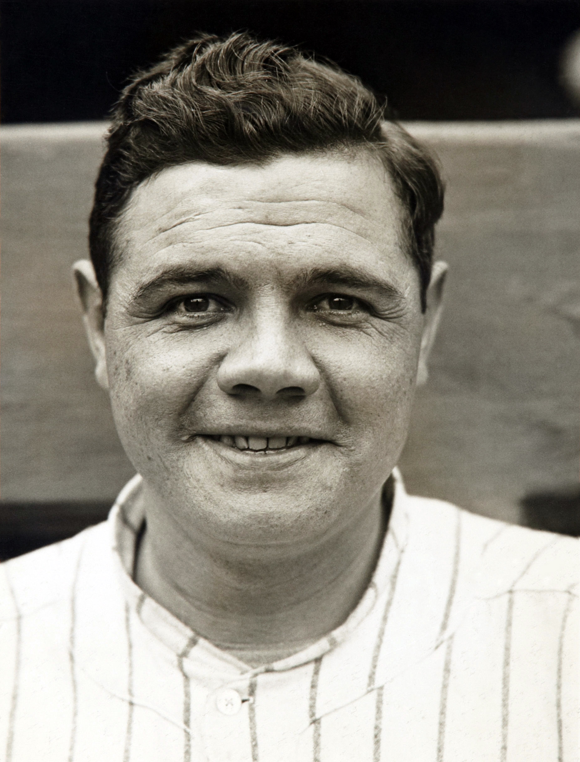
Many people don't know that Babe Ruth had severe amblyopia.

Many people don't know that Babe Ruth had severe amblyopia.

You probably know that George Herman Ruth Jr., also known as Babe, The Great Bambino, and The Sultan of Swat, was indisputably one of the greatest baseball players of all time. You may know that he came up with the Boston Red Sox before joining the New York Yankees. You might even know that began his career as a pitcher.

But did you know he had 20/200 vision in his left eye? Shocking, isn't it?

After his playing days were over, Ruth was examined by Dr. Gerald Kara, who found that the prolific slugger had surprisingly poor vision in his left eye (and 20/15 vision in his right), which Dr. Kara attributed to amblyopia. Because Ruth said he had never had an eye exam before, it is theoretically possible that his poor vision developed after his career was over, but Dr. Kara found no evidence that this was the case.

Amblyopia is the most common cause of reversible vision loss in children, affecting between 3-5% of all kids. It is defined as decreased vision due to abnormal development of vision in childhood, typically in one eye, and most commonly occurs due to unrecognized need for glasses or due to eye misalignment (strabismus). It is treated sometimes with glasses wear, and most frequently by patching the child's better-seeing eye. This forced use of the amblyopic eye helps the neural connections between the eye and brain develop properly, allowing for better vision.

I love treating amblyopia. Seeing a child start with one eye that doesn't see well at all, and then, over a period of months, thanks to the diligent work of the patient and parents in helping him/her keep the patch on, watching the vision improve -- it's a very gratifying experience.

With regard to Ruth, he is just one of the countless examples of people with a vision problem overcoming their circumstances and excelling in life.

Stars' Eyes: Jeremy Poincenot

Over the past year or so, on my Facebook and Instagram accounts, I've shared dozens of short posts with the tag "Stars Eyes Sunday," describing a famous person with an eye disease. These were popular posts, and I plan to continue sharing similar stories on this blog. Here's the first!

Jeremy Poincenot ("Points-en-oh") was a 19-year-old college sophomore at San Diego State when he suddenly lost his central vision in his right eye, followed shortly thereafter by his left. He was diagnosed with a rare optic nerve disorder, called Leber hereditary optic neuropathy (LHON).

LHON is a genetic condition caused by mutations in the DNA within mitochondria. Patients with LHON typically develop sudden, painless, central vision loss in one eye, followed weeks later by the other eye. The likelihood of developing LHON in a person who carries the mutation depends in part on what fraction of their mitochondria, the energy-producing organelles within each cell, harbor the genetic change. LHON occurs most frequently in young males, but females and much older patients can be affected, as well.

Jeremy hasn't let his severe vision loss hamper his love of the links. In fact, just today, he won the United States Blind Golf Association's National Championship! Jeremy has been featured on ABC's 20/20, MTV, and GolfWorld, and his website can be found here.

Jeremy Poincenot, winner of the 2016 US Blind Golf Championship, as shared on his Twitter account.

Jeremy Poincenot, winner of the 2016 US Blind Golf Championship, as shared on his Twitter account.

Will wearing glasses fix it?

Wearing glasses helps fix some eye problems, but certainly not all of them.

Wearing glasses helps fix some eye problems, but certainly not all of them.

There is a common misconception that any eye problem can be fixed by wearing the proper glasses. This, unfortunately, is not true. Glasses and contact lenses help correct eye problems related to focus, and that's it.

Think of the eye as a camera. If the camera is out of focus, then it won't take a good picture. Focus the camera (or wear glasses, in our analogy), and you'll have a clear image.

Extending the analogy, here are a few categories of eye problems that cannot be fixed by wearing glasses:

Cataract (lens smudged)
Corneal disease (cloudy screen)
Optic nerve disease (dead battery)
Retinal disease (bad film)